























Pancragen Peptide (20mg)
$63.00
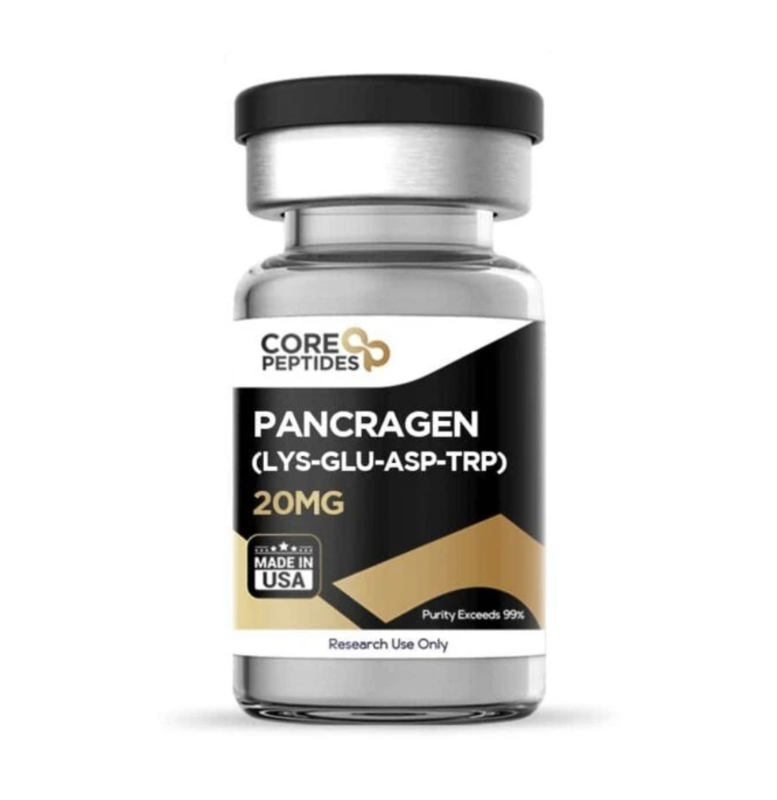
Pancragen peptide 20 mg provides a high-purity, laboratory-verified lyophilized peptide manufactured under strict quality control standards in the USA. Formulated with 20 mg of pure Pancragen—a synthetic tetrapeptide bioregulator composed of L-lysine, L-glutamic acid, L-aspartic acid, and L-tryptophan (Lys-Glu-Asp-Trp)—this compound is widely utilized in endocrinology, metabolic science, and cellular biology research examining pancreatic beta-cell restoration, insulin sensitivity modulation, and glucose homeostasis regulation.
Key Specifications
Active Compound: Pancragen (L-Lysyl-L-Glutamyl-L-Aspartyl-L-Tryptophan / Lys-Glu-Asp-Trp)
Chemical Formula: \text{C}_{26}\text{H}_{36}\text{N}_{6}\text{O}_{9}
Concentration: 20 mg per vial
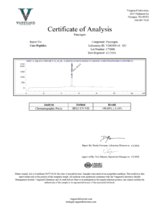
Purity Standard: Exceeds 99% (Third-Party HPLC Verified)
Formulation: Sterile Lyophilized Powder
Origin: Manufactured in USA
Classification: Research Use Only
Primary Research Applications
Pancreatic Beta-Cell Function & Insulin Sensitivity: Pancragen is extensively studied for its capacity to stimulate insulin gene expression, promote pancreatic islet cell functional integrity, and enhance peripheral glucose clearance in metabolic models.
Exocrine & Endocrine Tissue Remodeling: Evaluated in pancreatic insufficiency and pancreatitis models to assess its tissue-protective properties, acinar cell repair mechanisms, and digestive enzyme regulation.
Epigenetic Regulation of Carbohydrate Metabolism: Investigated in peptide bioregulator research for its interaction with chromatin structural proteins, regulating transcription factors tied to glycemic control and vascular protection against hyperglycemia.
Pancragen Peptide
Pancragen peptide 20mg is a tetrapeptide with the sequence Lys-Glu-Asp-Trp (KEDW). It is a synthetic structural analog of a peptide isolated from cattle pancreatic cells, which is posited to be a prospective peptide bioregulator for pancreatic function and aging-related metabolic problems. Pancragen has been suggested by researchers to act by penetrating cellular membranes and interacting with the nucleus and nucleolus. This suggests it could influence gene transcription associated with cell differentiation within the pancreatic cells. Key differentiation factors like Ptf1a, Pdx1, Pax6, Foxa2, Nkx2.2, and Pax4 are thought to play essential roles in the proper differentiation of pancreatic cells. Pancragen might up-regulate the expression of these factors, implying a positive action on the maturation of pancreatic cells. Pancragen may also influence the expression of molecules such as MMP2, MMP9, serotonin, CD79alpha, Mcl1, PCNA, and Ki67, suggesting it might boost the functional activity of pancreatic cells. Another mechanism may include a decrease in proapoptotic protein p53 and increase in antiapoptotic protein Mcl1 hinting at an antiapoptotic potential. Last but not least, Pancragen may influence aging biomarkers, reducing caspase-3 and cathepsin B activities and modulating levels of TNF-α and IGF-I. IGF-I, researched for its antiapoptotic actions, suggests that Pancragen’s actions might be linked to metabolic correction and antiapoptotic mechanisms. Pancragen also seems to affect the methylation patterns of genes like PDX1, PAX6, and NGN3, which might mediate some of its anti-aging potential on the pancreatic cells.
Chemical Makeup
Molecular formula: C26H36N6O9
Molecular weight: 576.25 g/mol
Sequence: Lys-Glu-Asp-Trp
Other known titles: KEDW, DWa (amidate)
Research and Clinical Studies
In the information below, we have broken down the latest clinical and preclinical research on Pancragen’s potential as observed in various experimental models.
Pancragen and the Pancreas
The tetrapeptide Pancragen has been studied for its potential impact on pancreatic cells, particularly in relation to cell differentiation and the regulation of insulin and glucagon release, crucial elements in the pancreatic cells’ endocrine function. (1)Research posited that Pancragen might penetrate cellular membranes to interact with the nucleus and nucleolus, thereby possibly influencing the transcription of genes crucial for cellular differentiation within the pancreatic gland. Key differentiation factors such as Ptf1a, Pdx1, Pax6, Foxa2, Nkx2.2, and Pax4 are crucial for the proper functioning and differentiation of various pancreatic cell types. It is posited that Pancragen’s interaction may potentially up-regulate the expression of these factors, which may play a role in the cellular maturation process of both acinar and islet cells in the pancreatic gland. The experiments conducted on embryonic cultures of pancreatic acinar cells in this study suggested that Pancragen might significantly enhance the expression of Ptf1a and Pdx1 proteins, which are known to be crucial for the maturation of acinar and islet cells. The study suggested that Pancragen’s actions might be more pronounced in aged cultures, where an apparent decrease in the expression of these proteins is observed as a part of the cellular aging process. This potential up-regulation of key differentiation factors by Pancragen is posited to possibly lead to an increased differentiation of pancreatic cells, and, subsequently, may aid in restoring the functional activity of the pancreatic cells to a state akin to that observed in younger cell cultures. The researchers also posited that “transcription factors that regulate differentiation of pancreatic cells are a pharmacological target for Pancragen, which allows considering it as an effective tool in the treatment of diabetes mellitus and pancreatitis.”
Another research study using murine models to gauge the impact of Pancragen on the pancreas’s functional morphology, also yielded interesting findings.(2)Initially, when DM was induced in murine models, there was a noted decrease in insulin-producing B cells and an uptick in glucagon-producing A cells. Such changes point to disrupted pancreatic cell functionality. However, upon exposure to Pancragen, potentially encouraging shifts were observed. Murine models showed evident compensatory changes in pancreatic cells and tissue. Specifically, Pancragen seemed to bolster insulin production from B cells while tempering glucagon production from A cells. Additionally, the proliferative activity of certain cells and their apoptosis seemed to normalize, closely resembling control murine models.
Further research has also suggested that Pancragen might play a pivotal role in the modulation of various cellular markers and proteins associated with the vitality of pancreatic cells. When applied to aging pancreatic cells, there was an observed increase in the expression of matrix metalloproteinase MMP2 and MMP9, serotonin, glycoprotein CD79alpha, the antiapoptotic protein Mcl1, and proliferation markers PCNA and Ki67. Conversely, there was a decrease in the expression of the proapoptotic protein p53 in aged pancreatic cell cultures. From these observations, it can be posited that the tetrapeptide potentially holds the capability to activate the expression of signaling molecules that serve as markers of the functional activity of pancreatic cells.(3)
Pancragen and Metabolic Problems
Studies have delved into the potential of Pancragen on metabolic problems in aged test subjects. For example, in one study, the focus was mainly on carbohydrate metabolism and the potential role Pancragen may play in regulating it.(4) The introduction of Pancragen was observed to significantly lower fasting glucose levels during a standard glucose tolerance test, alongside a reduction in insulin concentrations and the insulin resistance index. This suggests a potential utility of Pancragen in addressing the disturbances in carbohydrate metabolism, especially given the observed persistence of its glucose-lowering action post-exposure. It is critical to note that the study was focused on august test subjects, and the scope of Pancragen’s potential may be contingent on several other variables, including the severity of insulin resistance and other factors. The researchers also commented that “Administration of the tetrapeptide Pancragen is a promising approach to the correction of insulin resistance in elderly.”
Another study aimed to investigate the potential impact of Pancragen on the endocrine function of the pancreatic cells and the metabolic status of test models.(5) Aged test models used in this research were used to understand the potential of Pancragen in addressing age-related dysfunctions in the overall metabolism as well as the pancreatic islet apparatus. In older test models, there was a noted reduced rate of glucose utilization compared to younger counterparts. Moreover, higher insulin and C-peptide peaks were observed 5 and 15 minutes post-glucose introduction. However, when Pancragen was introduced per test model daily over 10 days, there was a marked improvement in the glucose utilization rate. This intervention also seemed to normalize the dynamics of plasma insulin and C-peptide in response to glucose. Intriguingly, the actions of Pancragen lingered, with some aspects of metabolic status and pancreatic cell function remaining improved even three weeks after the cessation of the trial.
In a study examining the actions of Pancragen on endothelial function within the setting of chronic hyperglycemia using murine models, it was observed that the introduction of Pancragen may have potential role in restoring endothelial adhesive characteristics. (6) Restoring these endothelial adhesive characteristics is crucial because, in models of metabolic problems, proper endothelial function plays a vital role in blood flow regulation and preventing complications such as atherosclerosis. Deficiencies or abnormalities in endothelial adhesion may increase susceptibility to vascular damage, making this restoration a key target for averting it.
Pancragen and Aging
Pancragen has been posited as a possible bioregulator for adjusting metabolic problems associated with aging. To understand Pancragen’s impact on aging, a study examined its potential on murine models across different age groups.(7) There was a noted decrease in certain aging biomarkers like caspase-3 and cathepsin B activities when Pancragen was introduced to the younger murine models. Interestingly, in the mature murine models, Pancragen application resulted in a significant reduction of TNF-α levels and an elevation in IGF-I, both of which are critical indicators related to aging and metabolic processes. The study also delved into a rapid experimental aging model by inducing diabetes mellitus in murine models. It was observed that Pancragen potentially normalized blood glucose levels, emphasizing its previously identified hypoglycemic properties. Furthermore, the presence of Pancragen might have suppressed certain apoptotic enzymatic components in the pancreatic cells, suggesting that its biological actions may be associated with both metabolic correction and antiapoptotic mechanisms. The actions of Pancragen on natural biological aging are potentially realized at the IGF-I level, known for its “survival factor” antiapoptotic actions. Moreover, the tetrapeptide’s potential interaction with certain genes suggests that its influence might extend to the proteolytic processing level.
According to another trial, Pancragen has been posited to tissue-specifically influence gene expression in pancreatic cell cultures.(8) The study suggests that variations in the methylation patterns of the PDX1, PAX6, and NGN3 gene promoter regions in pancreatic cells may be associated with aging and could potentially be the cause behind the alterations in their expression levels. This implies that long-standing changes in gene expression upon aging might be driven by modifications in these promoter methylation patterns. However, the expression levels of the PAX4 and FOXA2 genes in pancreatic cells seem to deviate during aging and in response to Pancragen, even when the methylation patterns of the PAX4 gene remain stable. Curiously, the FOXA2 gene’s promoter region in pancreatic cells exhibited only a handful of methylated CpG sites, with their methylation levels being influenced by both cell culture aging and Pancragen. Yet, this did not correlate straightforwardly with changes in gene expression levels. This suggests that, despite the influence of Pancragen on certain gene methylation patterns, the exact relationship between methylation and gene expression may be intricate and possibly regulated by other, yet unidentified, mechanisms.
References:
- Khavinson, V. K.h, Durnova, A. O., Polyakova, V. O., Tolibova, G. H., Linkova, N. S., Kvetnoy, I. M., Kvetnaia, T. V., & Tarnovskaya, S. I. (2013). Effects of pancragen on the differentiation of pancreatic cells during their ageing. Bulletin of experimental biology and medicine, 154(4), 501–504. https://doi.org/10.1007/s10517-013-1987-6
- Kvetnoi, I. M., Ryzhak, A. P., Kostyuchek, I. N., & Tafeev, Y. A. (2007). Effect of tetrapeptide pancragene on functional morphology of the pancreas in rats with experimental diabetes mellitus. Bulletin of experimental biology and medicine, 143(3), 368–371. https://doi.org/10.1007/s10517-007-0114-y
- Khavinson, V. K.h, Sevost’ianova, N. N., Durnova, A. O., Lin’kova, N. S., Tarnovskaia, S. I., Dudkov, A. V., & Kvetnaia, T. V. (2012). Advances in gerontology = Uspekhi gerontologii, 25(4), 680–684.
- Korkushko, O. V., Khavinson, V. K.h, Shatilo, V. B., Antonyk-Sheglova, I. A., & Bondarenko, E. V. (2011). Prospects of using pancragen for correction of metabolic disorders in elderly people. Bulletin of experimental biology and medicine, 151(4), 454–456. https://doi.org/10.1007/s10517-011-1354-4


Related products
ACE-031 Peptide (1mg)
ACE-031 1 mg is a research-grade lyophilized protein peptide engineered for high-purity laboratory applications. Each vial contains 1 mg of HPLC-verified ACE-031, third-party tested to guarantee over 99% purity and manufactured in the USA under strict standards. Formulated specifically for research investigating myostatin signal neutralization, ActRIIB decoy receptor binding, and muscle growth regulation pathways, this product is strictly intended for research use only.
AICAR Peptide (50mg)
AICAR 50 mg provides a high-purity, laboratory-verified lyophilized compound manufactured under strict quality control standards in the USA. Formulated with 50 mg of pure AICAR (5-Aminoimidazole-4-carboxamide ribonucleotide) per vial, this synthetic adenosine analog is widely utilized in cellular, metabolic, and exercise physiology research investigating AMP-activated protein kinase (AMPK) activation, cellular energy homeostasis, and mitochondrial biogenesis pathways.
Key Specifications
Active Compound: AICAR (Acadesine / AICA Riboside)
Chemical Formula: \text{C}_{9}\text{H}_{15}\text{N}_{4}\text{O}_{8}\text{P}
Concentration: 50 mg per vial
Purity Standard: Exceeds 99% (Third-Party HPLC Verified)
Formulation: Sterile Lyophilized Powder
Origin: Manufactured in USA
Classification: Research Use Only
Primary Research Applications
AMP-Activated Protein Kinase (AMPK) Activation: AICAR is intracellularly phosphorylated into ZMP, an AMP mimic that directly activates AMPK without altering cellular ATP/ADP ratios, facilitating targeted metabolic research.
Mitochondrial Biogenesis & Endurance Pathways: Applied in cell cultures to analyze upregulated PGC-1$\alpha$ signaling, glucose uptake enhancement, and lipid oxidation mechanisms.
Ischemic & Cellular Energy Homeostasis Studies: Extensively investigated in cardiac and skeletal muscle models measuring metabolic adaptation, cellular stress response, and insulin sensitivity.
B7-33 (6mg)
B7-33 6 mg provides a high-purity, laboratory-verified lyophilized peptide manufactured under strict quality control standards in the USA. Formulated with 6 mg of pure B7-33—a single-chain synthetic peptide functional analog of human gene-2 relaxin (H2 relaxin)—this compound is widely utilized in cardiovascular, nephrology, and matrix biology research examining selective RXFP1 receptor signaling, extracellular matrix (ECM) remodeling, and anti-fibrotic signaling pathways without inducing tumor growth pathways.
Key Specifications
Active Compound: B7-33 (Relaxin-2 Receptor Agonist)
Chemical Formula: \text{C}_{136}\text{H}_{210}\text{N}_{40}\text{O}_{41}
Concentration: 6 mg per vial
Purity Standard: Exceeds 99% (Third-Party HPLC Verified)
Formulation: Sterile Lyophilized Powder
Origin: Manufactured in USA
Classification: Research Use Only
Primary Research Applications
Targeted Anti-Fibrotic Activity: B7-33 acts through the RXFP1 receptor to upregulate matrix metalloproteinases (MMPs) and suppress excessive collagen deposition, providing a targeted model for reversal of organ fibrosis.
Cardiovascular & Renal Remodeling: Extensively evaluated in preclinical models of heart failure, renal scarring, and vascular stiffness to assess hemodynamic regulation and arterial compliance enhancement.
Biasing RXFP1 Receptor Signaling: Investigated for its ability to preferentially stimulate pERK signaling over cAMP accumulation, preventing unwanted inotropic or proliferative side effects associated with full-length native relaxin.
Epithalon (25mg)
Epithalon 25 mg is a research-grade lyophilized tetrapeptide engineered for high-purity laboratory applications. Each vial contains 25 mg of HPLC-verified Epithalon, third-party tested to guarantee over 99% purity and manufactured in the USA under strict standards. Formulated specifically for research investigating telomerase enzyme induction, pineal function restoration, and cellular longevity pathways, this product is strictly intended for research use only.
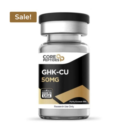
GHK-Cu (Copper) (50mg)
GHK-Cu (50mg) is a premium copper-binding tripeptide engineered to support advanced tissue repair, collagen synthesis, and cellular regeneration. Naturally present in human plasma, GHK-Cu levels decline with age. This potent peptide actively stimulates collagen and elastin production, accelerates wound healing, reduces fine lines, and promotes skin firmness and hair follicle restoration. Ideal for anti-aging and regenerative research applications.
Key Benefits: Boosts collagen & elastin, aids tissue remodeling, enhances skin barrier health, supports hair density.
Format: 50mg lyophilized powder (purity ≥99%).
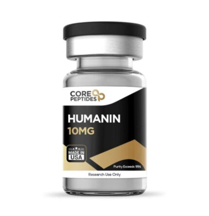
Humanin (10mg)
Humanin 10 mg provides a high-purity, laboratory-verified lyophilized peptide manufactured under strict quality control standards in the USA. Formulated with 10 mg of pure Humanin—a naturally occurring 24-amino-acid micropeptide encoded within the mitochondrial genome (16S rRNA)—this compound is widely utilized in cellular biology, metabolic health, and neurodegenerative disease research investigating cytoprotective mechanisms, oxidative stress regulation, and insulin sensitivity pathways.
Key Specifications
Active Compound: Humanin (HN)
Chemical Formula: \text{C}_{119}\text{H}_{204}\text{N}_{34}\text{O}_{35}\text{S}_{2}
Concentration: 10 mg per vial
Purity Standard: Exceeds 99% (Third-Party HPLC Verified)
Formulation: Sterile Lyophilized Powder
Origin: Manufactured in USA
Classification: Research Use Only
Primary Research Applications
Cytoprotection & Anti-Apoptotic Pathways: Humanin is studied for its ability to suppress Bax-mediated apoptosis, protecting various cell lines against programmed cell death triggered by toxic insults.
Neuroprotective Dynamics: Extensively evaluated in Alzheimer’s disease and neurodegenerative models to analyze its role in mitigating amyloid-beta (A\beta) cytotoxicity and preserving neuronal function.
Mitochondrial & Metabolic Homeostasis: Investigated for its regulatory actions on insulin sensitivity, glucose metabolism, and cellular homeostasis under conditions of metabolic stress.
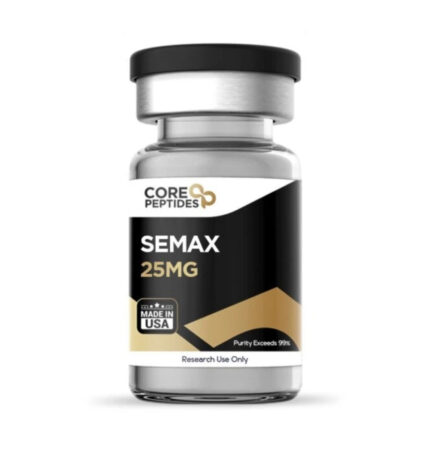
Semax (25mg)
Semax (25mg) is a premium research-grade synthetic heptapeptide (≥98% purity) studied for its potent neuroprotective, memory-enhancing, and BDNF-boosting properties. Supplied as a stable, lyophilized powder, every batch is HPLC and Mass Spectrometry verified to ensure maximum purity and consistency for laboratory investigation.
For research and laboratory use only. Not for human consumption.
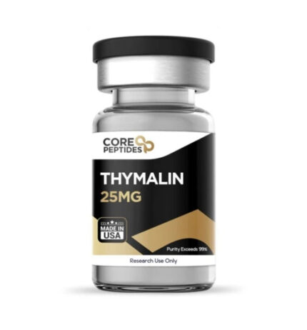
Thymalin Peptide (25mg)
Thymalin peptide 25 mg provides a high-purity, laboratory-verified lyophilized peptide manufactured under strict quality control standards in the USA. Formulated with 25 mg of pure Thymalin—a synthetic bioregulator peptide complex derived from thymic polypeptide fraction research—this compound is widely utilized in immunology, endocrinology, and biogerontology research examining T-lymphocyte differentiation, immune system homeostasis, and neuroendocrine-immune interactions.
Key Specifications
Active Compound: Thymalin (Thymic Peptide Bioregulator)
Chemical Structure: Synthetic Thymic Dipeptide Complex (Lys-Glu / Glu-Trp analogs)
Concentration: 25 mg per vial
Purity Standard: Exceeds 99% (Third-Party HPLC Verified)
Formulation: Sterile Lyophilized Powder
Origin: Manufactured in USA
Classification: Research Use Only
Primary Research Applications
Immunomodulation & T-Cell Maturation: Thymalin is studied for its ability to regulate T-lymphocyte differentiation, restore helper/suppressor T-cell ratios (CD4^+/CD8^+), and stimulate cellular immunity markers.
Thymic Involution & Immune Restoration: Evaluated in age-related immune dysfunction and immunosenescence models to measure restored thymic secretory capacity and cytokine balance.
Neuroendocrine-Immune Axis Regulation: Extensively investigated in peptide bioregulator research for its role in normalizing neuroendocrine response, pineal-thymus interaction, and gene expression pathways during chronic stress or pathology.
Reviews
There are no reviews yet.