






















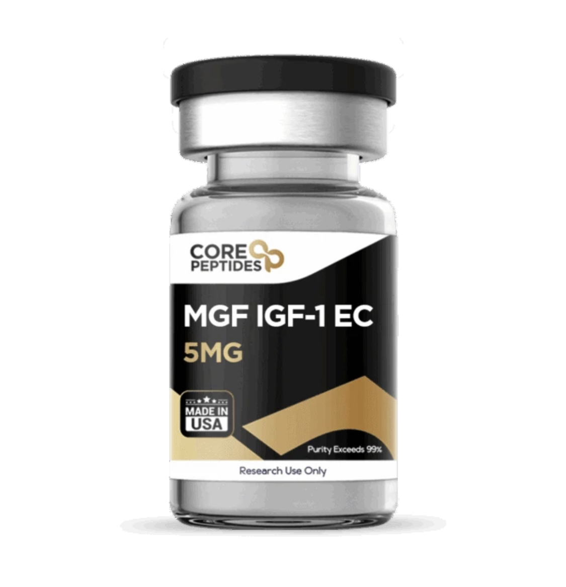
MGF IGF-1 EC (5mg)
$140.00
MGF IGF-1 EC 5 mg provides a high-purity, laboratory-verified lyophilized peptide manufactured under strict quality control standards in the USA. Formulated with 5 mg of pure Mechano-Growth Factor (MGF, a splice variant of Insulin-like Growth Factor 1 designated as IGF-1EC in humans)—this naturally expressed splice variant is generated in response to mechanical overload and muscle tissue strain. It is widely utilized in exercise physiology, musculoskeletal biology, and regenerative medicine research examining satellite cell activation, local tissue hypertrophy, and accelerated muscular repair mechanisms.
Key Specifications
Active Compound: MGF (Mechano-Growth Factor / IGF-1EC)
Chemical Formula: \text{C}_{121}\text{H}_{200}\text{N}_{42}\text{O}_{39}
Concentration: 5 mg per vial
Purity Standard: Exceeds 99% (Third-Party HPLC Verified)
Formulation: Sterile Lyophilized Powder
Origin: Manufactured in USA
Classification: Research Use Only
Primary Research Applications
Satellite Cell Activation & Myoblast Proliferation: MGF (IGF-1EC) acts as a primary local signal following mechanical tissue stress, stimulating quiescent muscle satellite cell activation, proliferation, and pool expansion.
Skeletal Muscle Hypertrophy & Repair: Extensively studied in sports science and orthopedics for its localized anabolic action, accelerating myotube formation and protein synthesis without systemic IGF-1 receptor activation.
Cardioprotection & Neuroprotection: Evaluated in cardiac and neurological injury models to examine its potential in preventing cellular apoptosis, reducing ischemic tissue damage, and supporting localized repair cascades.
MGF IGF-1 Ec Peptide
MGF IGF-1 Ec is the Mehano Growth Factor (MGF) domain made of 24 amino acids, which have been cleaved from the 110 amino acids of IGF-1 Ec alongside other important molecules such as IGF-1 (70 amino acids). Thus, MGF IGF-1 Ec corresponds to the C-terminal 24 residues of the IGF-IEb/Ec pro-peptide.
According to research by Janssen et al, the peptide fragment does not measurably induce IGF-1 receptor tyrosine phosphorylation, and does not activate insulin receptors.(1)Instead, the peptide appears to have potential to function as an IGF-related peptide that bypasses classical IGF-1 receptor activation and instead may signal preferentially through ERK pathways, exerting an anabolic potential towards a variety of cell cultures, especially muscle cells.
Chemical Makeup
Other Known Titles: Mechano-Growth Factor, MGF-Ct24E
Molecular Weight: 2971.99 g/mol
Molecular Formula: C124H204N42O41S1
Research and Clinical Studies
MGF IGF-1 Ec and Muscle Cell Hyperthrophy
Most of the research on MGF IGF-1 Ec fragments revolved around muscle cells. One of the most important experiments on the peptide was conducted by Li et al., and suggests that the peptide apparently supports muscle cell hypertrophy.(2) Interestingly, it appears to achieve that by activating Erk5 and Erk1/2 while only weakly and transiently engaging Akt, and it may do so without detectable IGF-1 receptor tyrosine phosphorylation. This profile suggests potential implications of the peptide in studies conducted in laboratory settings aiming to dissect noncanonical, Erk5-centered pathways downstream of IGF-1-derived products, separate from the typical IGF-1–Akt proliferative axis.
Li et al. also suggest that in cultured intestinal smooth muscle cells, the peptide may promote an increase in cell volume and total protein content, without stimulating DNA synthesis. This contrasts with IGF-1, which drives proliferation and reduces average cell volume. Because the researchers comment that “Erk5 [mitigation] or MEF2C siRNA blocked smooth muscle-specific gene expression and hypertrophy induced by synthetic MGF”, the peptide may support laboratory study Erk5–MEF2C–dependent transcriptional programs. Future research may interact with the peptide for studying smooth muscle–specific genes such as α-smooth muscle actin, γ-actin, smoothelin, and desmin, all of which appear upregulated by MGF IGF-1 Ec in a MEF2C-dependent manner.
MGF IGF-1 Ec and Muscle Cell Aging
Kandalla et al. also suggest that MGF IGF-1 Ec may act as a modulator of cellular aging in muscle cells. In myoblasts derived from young satellite cells, repeated short exposures to MGF IGF-1 Ec seemed to mitigate cellular aging for longer in the cell cultures observed in the study.(3) The exposed cells were able to go through slightly more rounds of division, and, importantly, a larger share of them were still actively dividing even after many cycles of growth. In contrast, the control cells largely stopped dividing at that stage.
These findings fit with the idea that MGF IGF-1 Ec may briefly interact with the cell-cycle machinery in a way that is at least partly similar to IGF-1 and may interact with stress-response pathways that control when cells permanently stop dividing. However, the authors stress that any interaction with cellular lifespan is modest rather than dramatic.
In older muscle cells, MGF IGF-1 Ec did not prolong the period during which cells may keep dividing and may even have accelerated the decline in their division activity. This cellular age-dependent difference in response suggests that MGF IGF-1 Ec may be a helpful tool for experimentally comparing more aged cells versus less aged cellular senescence programs under the same culture conditions.
MGF IGF-1 Ec and Muscle Cell Fibrosis
Another team of researchers led by Liu et al. suggests that the peptide may reduce histological fibrosis and lower early expression of collagen I and III.(4) This suggests the peptide may have an anti-fibrotic potential in injured muscle cells. In parallel, MGF IGF-1 Ec appeared to decrease “the expression of muscle [cell] inflammatory cytokines (TNF-α, IFN-γ, IL-1β, and TGF-β), chemokines (CCL2, CCL5, and CXCR4), oxidative stress factors (gp91phox) and matrix metalloproteinases (MMP-1, MMP-2, MMP-9, MMP-10, and MMP-14)”, thus hinting at diminished NADPH oxidase–related oxidative stress.
The researchers suggested that the peptide may have also altered the expression of several MMPs involved in extracellular matrix turnover. Taken together, the research by Liu et al suggests that the MGF IGF-1 Ec fragment may prove to be a relevant laboratory tool to investigate and possibly modulate the balance between muscular tissue repair and fibrotic scarring.
MGF IGF-1 Ec and Muscle Cell Death
Experiments by Doroudian et al. have also explored MGF IGF-1 Ec as a potential modulator of cellular death.(5) According to researchers, the anti-apoptotic potential of the peptide was clear in the mammalian myocyte cultures exposed to low oxygen environments observed in laboratory settings. The researchers observed less DNA fragmentation and increased expression of the pro-survival gene Bcl-2 compared with hypoxic control cells. These findings suggest that the peptide may prove to be relevant experimentally as a tool to probe survival pathways in muscle cells under controlled stress.
Mechanistically, the upregulation of Bcl-2 suggests a possible engagement of intrinsic survival signaling, likely intersecting with mitochondrial pathways. Thus, the data position MGF IGF-1 Ec as a potential research reagent for reducing apoptosis in stressed muscle cells and for systematically studying how IGF-1–related peptides may promote cell survival.
MGF IGF-1 Ec and Cartilage Cells
In further research, the team of Liu et al. also investigated the potential of the MGF IGF-1 Ec fragment in other cell cultures, such as cartilage cells.(6) The data summarized by the researchers suggests that MGF IGF-1 Ec may support several key processes around defect zones that form when cartilage cells are mechanically overloaded or otherwise stressed.
In progenitor cell cultures, the peptide apparently potentiated TGF-β3–induced chondrogenesis, supporting Col2 and aggrecan expression while suppressing Col1. This suggests that the peptide may be a possible cofactor in systems that model matrix quality, thus stimulating hyaline rather than fibrocartilage-like cellular production. In damaged or hypoxic chondrocyte cultures, the peptide was suggested to reduce pro-fibrotic and catabolic markers (Col1, MMP1/3/13, HIF-1α) and to increase Col2, often via PI3K–Akt and MEK–ERK1/2 signaling.
The researchers posited that MGF IGF-1 Ec may also act as a factor that promotes migration of chondrocytes and mesenchymal cells under overload or severe hypoxia, apparently through RhoA–YAP activation and associated focal adhesion and cytoskeletal reorganization. This gives it potential relevance in assays of cell motility, cytoskeletal mechanics, and mechanotransduction.
Some of the data analyzed by the researchers also suggest that MGF IGF-1 Ec may dampen inflammatory signaling by apparently downregulating IL-1β, TNF-α, and TGF-β. Moreover, the peptide may mitigate apoptosis by shifting Bcl-2/Bax balance and reducing caspase-3/-8 and CHOP, making it a candidate reagent for dissecting the potential interactions between mechanical stress, ER stress/UPR, and programmed cell death in cartilage cell cultures.
MGF IGF-1 Ec and Bone Cells
Research by Deng et al, also suggests that MGF IGF-1 Ec may act as a potential osteoanabolic signal with a distinct profile from IGF-1.(7)The researchers conducted experiments with MC3T3-E1 osteoblast-like cells, and noted that the peptide may stimulate proliferation more strongly than full-length MGF or IGF-1, with ~1.4-fold higher pro-proliferative activity than IGF-1. Mechanistically, the peptide may push cells into S and G2/M phases, doubling the number of cells synthesizing DNA and increasing the number of cells entering mitosis about 5-fold.
This potential was coupled with robust activation of the MAPK/ERK1/2 pathway. Once again, the researchers observed that blocking ERK almost abolished the proliferative response, whereas PI3K mitigation had minimal meaningful interaction, highlighting that MGF IGF-1 Ec may act via an ERK-dominant, IGF-1 receptor-independent mechanism.
Further laboratory research suggested that the peptide may increase callus formation, cortical bridging, and disappearance of the fracture line, with a subset of defects radiographically united by week 8. Histologically, the research models appeared to have increased levels of lamellar bone, restored marrow cavity, higher vascularity and osteoid formation, and fewer fibroblasts compared with controls—consistent with faster, more organized bone remodeling rather than fibrous non-union.
With this in mind, the peptide may be viewed as a selective supporter of osteoblast proliferation and early bone regeneration, acting through MAPK/ERK signaling and offering a compact, synthetic tool to model or potentially augment bone cell regeneration in laboratory systems.
MGF IGF-1 Ec and Neural-like Cells
Research by Quesada et al. suggests that MGF IGF-1 Ec may support cell survival, reduce apoptotic markers, and maintain mitochondrial integrity in SH-SY5Y neuroblastoma cells found in mammalian models with mitochondrial dysfunction and increased apoptosis due to neurotoxin exposure.
Mechanistically, this interaction has been linked to upregulation of heme-oxygenase-1 (HO-1), which, when mitigated, appears to negate the peptide’s protective potential. Further research has similarly suggested that MGF IGF-1 Ec may reduce loss of vulnerable neuronal populations and preserve motor performance under neurotoxic or degenerative conditions. Moreover, the peptide may mimic a stress-protective, ERK-linked stimulus in neural-like cells.


Related products
B7-33 (6mg)
B7-33 6 mg provides a high-purity, laboratory-verified lyophilized peptide manufactured under strict quality control standards in the USA. Formulated with 6 mg of pure B7-33—a single-chain synthetic peptide functional analog of human gene-2 relaxin (H2 relaxin)—this compound is widely utilized in cardiovascular, nephrology, and matrix biology research examining selective RXFP1 receptor signaling, extracellular matrix (ECM) remodeling, and anti-fibrotic signaling pathways without inducing tumor growth pathways.
Key Specifications
Active Compound: B7-33 (Relaxin-2 Receptor Agonist)
Chemical Formula: \text{C}_{136}\text{H}_{210}\text{N}_{40}\text{O}_{41}
Concentration: 6 mg per vial
Purity Standard: Exceeds 99% (Third-Party HPLC Verified)
Formulation: Sterile Lyophilized Powder
Origin: Manufactured in USA
Classification: Research Use Only
Primary Research Applications
Targeted Anti-Fibrotic Activity: B7-33 acts through the RXFP1 receptor to upregulate matrix metalloproteinases (MMPs) and suppress excessive collagen deposition, providing a targeted model for reversal of organ fibrosis.
Cardiovascular & Renal Remodeling: Extensively evaluated in preclinical models of heart failure, renal scarring, and vascular stiffness to assess hemodynamic regulation and arterial compliance enhancement.
Biasing RXFP1 Receptor Signaling: Investigated for its ability to preferentially stimulate pERK signaling over cAMP accumulation, preventing unwanted inotropic or proliferative side effects associated with full-length native relaxin.
BPC-157 (10 mg)
BPC-157 (10mg) is a high-purity (≥98%) research-grade pentadecapeptide extensively studied for its powerful tissue regeneration, tendon healing, and gut mucosa protective properties. Supplied as a stable lyophilized powder, every batch undergoes strict HPLC and Mass Spectrometry testing to guarantee maximum purity and reliable research results.
For research and laboratory use only. Not for human consumption.
Epithalon (25mg)
Epithalon 25 mg is a research-grade lyophilized tetrapeptide engineered for high-purity laboratory applications. Each vial contains 25 mg of HPLC-verified Epithalon, third-party tested to guarantee over 99% purity and manufactured in the USA under strict standards. Formulated specifically for research investigating telomerase enzyme induction, pineal function restoration, and cellular longevity pathways, this product is strictly intended for research use only.
GHK-Cu (Copper) (50mg)
GHK-Cu (50mg) is a premium copper-binding tripeptide engineered to support advanced tissue repair, collagen synthesis, and cellular regeneration. Naturally present in human plasma, GHK-Cu levels decline with age. This potent peptide actively stimulates collagen and elastin production, accelerates wound healing, reduces fine lines, and promotes skin firmness and hair follicle restoration. Ideal for anti-aging and regenerative research applications.
Key Benefits: Boosts collagen & elastin, aids tissue remodeling, enhances skin barrier health, supports hair density.
Format: 50mg lyophilized powder (purity ≥99%).
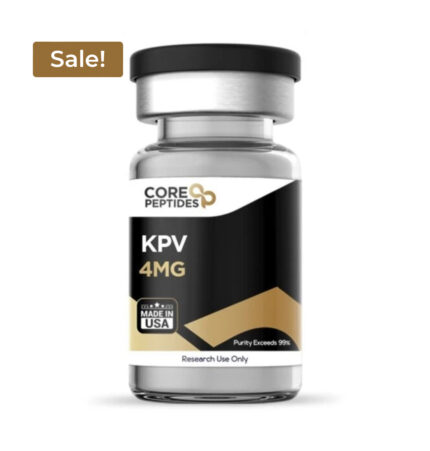
KPV Peptide (4mg)
KPV Peptide 4 mg provides a high-purity, laboratory-verified lyophilized peptide manufactured under strict quality control standards in the USA. Formulated with 4 mg of pure KPV (Lysine-Proline-Valine)—the C-terminal tripeptide fragment of alpha-melanocyte-stimulating hormone (\alpha\text{-MSH})—this compound is widely utilized in gastroenterology, immunology, and dermatology research investigating inflammatory bowel disease (IBD) suppression, antimicrobial signaling, and NF-\kappa\text{B} pathway downregulation.
Key Specifications
Active Compound: KPV (Lysine-Proline-Valine / \alpha\text{-MSH}_{11-13})
Chemical Formula: \text{C}_{16}\text{H}_{30}\text{N}_{4}\text{O}_{4}
Concentration: 4 mg per vial
Purity Standard: Exceeds 99% (Third-Party HPLC Verified)
Formulation: Sterile Lyophilized Powder
Origin: Manufactured in USA
Classification: Research Use Only
Primary Research Applications
Gastrointestinal Anti-Inflammatory Modulation: KPV is extensively studied in intestinal inflammation models for its ability to penetrate mucosal epithelial cells and inhibit nuclear factor kappa B (NF-\kappa\text{B}) translocation, reducing colonic tissue damage.
Antimicrobial & Anti-Fungal Activity: Evaluated in cellular microbiology models for direct antimicrobial properties against pathogen strains, including Candida albicans and Staphylococcus aureus.
Dermatological & Tissue Repair: Investigated in wound healing and inflammatory skin condition models to suppress pro-inflammatory cytokine expression (\text{IL-6}, \text{TNF-}\alpha) without inducing melanogenesis.
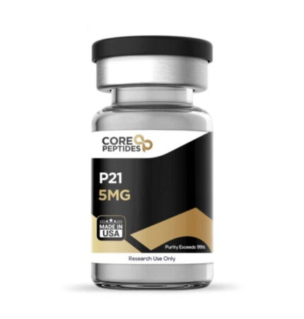
P21 Peptide (5mg)
P21 5 mg provides a high-purity, laboratory-verified lyophilized peptide manufactured under strict quality control standards in the USA. Formulated with 5 mg of pure P21—a synthetic peptide mimetic derived from ciliary neurotrophic factor (CNTF)—this compound is widely utilized in neurobiology, cognitive science, and neurodegenerative disease research examining neurogenesis, synaptic plasticity, BDNF signaling, and cognitive enhancement mechanisms.
Key Specifications
Active Compound: P21 Peptide (CNTF-derived synthetic peptide)
Chemical Formula: \text{C}_{30}\text{H}_{55}\text{N}_{9}\text{O}_{9}
Concentration: 5 mg per vial
Purity Standard: Exceeds 99% (Third-Party HPLC Verified)
Formulation: Sterile Lyophilized Powder
Origin: Manufactured in USA
Classification: Research Use Only
Primary Research Applications
Neurogenesis & Synaptogenesis: P21 is evaluated for its ability to promote neural progenitor cell proliferation and maturation in the dentate gyrus of the hippocampus, supporting research into structural brain plasticity.
Neurotrophic Factor Expression: Extensively studied for enhancing endogenous brain-derived neurotrophic factor (BDNF) and nerve growth factor (NGF) signaling pathways without inducing CNTF-receptor autoantibodies.
Cognitive Enhancement & Neuroprotection: Investigated in animal models of cognitive impairment, Alzheimer's disease, and traumatic brain injury to analyze memory retention recovery and synaptic density preservation.
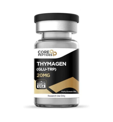
Thymagen Peptide (20mg)
Thymagen 20 mg provides a high-purity, laboratory-verified lyophilized peptide manufactured under strict quality control standards in the USA. Formulated with 20 mg of pure Thymagen—a synthetic dipeptide bioregulator consisting of L-glutamic acid and L-tryptophan (Glu-Trp)—this compound is widely utilized in immunology, molecular biology, and epigenetic research examining T-cell activation, chromatin structure modification, and immune system homeostasis regulation.
Key Specifications
Active Compound: Thymagen (L-Glutamyl-L-Tryptophan / Glu-Trp)
Chemical Formula: \text{C}_{16}\text{H}_{19}\text{N}_{3}\text{O}_{5}
Concentration: 20 mg per vial
Purity Standard: Exceeds 99% (Third-Party HPLC Verified)
Formulation: Sterile Lyophilized Powder
Origin: Manufactured in USA
Classification: Research Use Only
Primary Research Applications
Immune Response Modulation: Thymagen is extensively evaluated for its ability to regulate cellular immune activity, restore suppressed T-lymphocyte proliferation, and balance helper-to-suppressor T-cell ratios.
Epigenetic & Gene Expression Regulation: Studied in cell biology models for its capacity to interact with histone proteins and DNA sequences, influencing gene expression associated with protein synthesis and immune cell differentiation.
Tissue Regeneration & Stress Resistance: Investigated for its protective effects on hemopoiesis, resistance to oxidative injury, and systemic recovery mechanisms following toxic or infectious immune suppression.
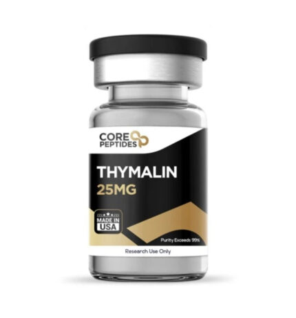
Thymalin Peptide (25mg)
Thymalin peptide 25 mg provides a high-purity, laboratory-verified lyophilized peptide manufactured under strict quality control standards in the USA. Formulated with 25 mg of pure Thymalin—a synthetic bioregulator peptide complex derived from thymic polypeptide fraction research—this compound is widely utilized in immunology, endocrinology, and biogerontology research examining T-lymphocyte differentiation, immune system homeostasis, and neuroendocrine-immune interactions.
Key Specifications
Active Compound: Thymalin (Thymic Peptide Bioregulator)
Chemical Structure: Synthetic Thymic Dipeptide Complex (Lys-Glu / Glu-Trp analogs)
Concentration: 25 mg per vial
Purity Standard: Exceeds 99% (Third-Party HPLC Verified)
Formulation: Sterile Lyophilized Powder
Origin: Manufactured in USA
Classification: Research Use Only
Primary Research Applications
Immunomodulation & T-Cell Maturation: Thymalin is studied for its ability to regulate T-lymphocyte differentiation, restore helper/suppressor T-cell ratios (CD4^+/CD8^+), and stimulate cellular immunity markers.
Thymic Involution & Immune Restoration: Evaluated in age-related immune dysfunction and immunosenescence models to measure restored thymic secretory capacity and cytokine balance.
Neuroendocrine-Immune Axis Regulation: Extensively investigated in peptide bioregulator research for its role in normalizing neuroendocrine response, pineal-thymus interaction, and gene expression pathways during chronic stress or pathology.
Reviews
There are no reviews yet.